Posts

Showing posts with the label X-Ray

Chest X Ray - Four Patterns

Image
Chest X Ray - Four Patterns Lung abnormalities with an increased density - also called opacities - are the most common. A practical approach is to divide these into four patterns: Consolidation Interstitial Nodules or masses Atelectasis  Whenever you see an area of increased density within the lung, it must be the result of one of these four patterns.   Consolidation   - any pathologic process that fills the alveoli with fluid, pus, blood, cells (including tumor cells) or other substances resulting in lobar, diffuse or multifocal ill-defined opacities.   Interstitial   - involvement of the supporting tissue of the lung parenchyma resulting in fine or coarse reticular opacities or small nodules.   Nodule or mass   - any space occupying lesion either solitary or multiple.   Atelectasis   - collapse of a part of the lung due to a decrease in the amount of air in the alveoli resulting in volume loss and increased den...

Air bronchogram

Image
What is  Air bronchogram Air bronchogram  refers to the phenomenon of  air -filled bronchi (dark) being made visible by the opacification of surrounding alveoli (grey/white). It is almost always caused by a pathologic airspace/alveolar process, in which something other than  air  fills the alveoli.

Consolidation

Image
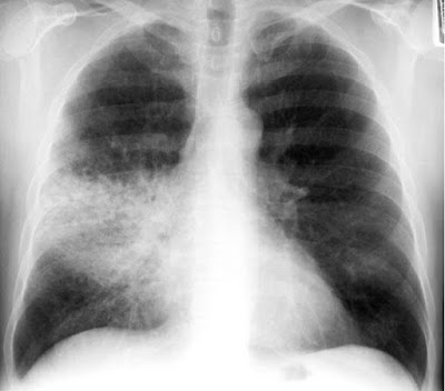
Consolidation The lung is said to be consolidated when the alveoli and small airways are filled with dense material. This dense material may consist of: •  Pus (pneumonia) •  Fluid (pulmonary oedema) •  Blood (pulmonary haemorrhage) •  Cells (cancer) Lobar consolidation: – Alveolar space filled with inflammatory exudate – Interstitium and architecture remain intact – The airway is patent – Radiologically: •  A density corresponding to a segment or lobe •  Air bronchogram, and •  No significant loss of lung volume

Air Bronchogram

Image
Air Bronchogram A tubular outline of an airway made visible due to the filling of the surrounding alveoli by fluid or inflammatory exudates. Conditions in which air bronchograms are seen: •  Lung consolidation •  Pulmonary oedema •  Non-obstructive pulmonary atelectasis •  Interstitial disease •  Neoplasm •  Normal expiration

Silhouette Sign

Image
Silhouette Sign •  The loss of the lung/soft tissue interface due to the presence of fluid in the normally air-filled lung •  If an intrathoracic opacity is in anatomic contact with a border, then the opacity will obscure that  border •  Commonly seen with the borders of the heart, aorta, chest wall, and diaphragm