Posts

Showing posts with the label Clinical Exam

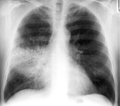
Air bronchogram

Image
What is  Air bronchogram Air bronchogram  refers to the phenomenon of  air -filled bronchi (dark) being made visible by the opacification of surrounding alveoli (grey/white). It is almost always caused by a pathologic airspace/alveolar process, in which something other than  air  fills the alveoli.

Bacterial Conjunctivitis Vs other causes

Image
Bacterial Conjunctivitis Vs other causes A clinical scoring system has been developed to distinguish bacterial from other causes of conjunctivitis in healthy adults who did not wear contact lenses.  A score of +5 to −3 is determined as follows: ~ Two glued eyes (+5); one glued eye (+2); history of conjunctivitis (−2); eye itching (−1). ~ A score of +5, +4, or +3 is useful in ruling in bacterial conjunctivitis with specificities of 100%, 94%, and 92%, respectively. ~ Scores of −1, −2, or −3 are useful in ruling out bacterial conjunctivitis with sensitivities of 98%, 98%, and 100%,  respectively. Allergic conjunctivitis is typically bilateral and accompanied by eye itching. Giant papillary conjunctivitis is a type of allergic reaction, most commonly to soft contact lenses

Consolidation

Image
Consolidation The lung is said to be consolidated when the alveoli and small airways are filled with dense material. This dense material may consist of: •  Pus (pneumonia) •  Fluid (pulmonary oedema) •  Blood (pulmonary haemorrhage) •  Cells (cancer) Lobar consolidation: – Alveolar space filled with inflammatory exudate – Interstitium and architecture remain intact – The airway is patent – Radiologically: •  A density corresponding to a segment or lobe •  Air bronchogram, and •  No significant loss of lung volume

Air Bronchogram

Image
Air Bronchogram A tubular outline of an airway made visible due to the filling of the surrounding alveoli by fluid or inflammatory exudates. Conditions in which air bronchograms are seen: •  Lung consolidation •  Pulmonary oedema •  Non-obstructive pulmonary atelectasis •  Interstitial disease •  Neoplasm •  Normal expiration

Silhouette Sign

Image
Silhouette Sign •  The loss of the lung/soft tissue interface due to the presence of fluid in the normally air-filled lung •  If an intrathoracic opacity is in anatomic contact with a border, then the opacity will obscure that  border •  Commonly seen with the borders of the heart, aorta, chest wall, and diaphragm

Monitoring glucose Levels in DM

Image
Monitoring Glucose Levels in DM • HbA 1c gives an estimate of the degree of glucose control over 2 to 3 months. • The American Diabetes Association recommends a treatment goal of HbA 1c <7.0%. In general, HbA 1c >10% is poor control, 8.5% to 10% is fair control, 7.0% to 8.5% is good control, and <7.0% is ideal. • The American Diabetes Association recommends keeping fasting blood glucose level <130 mg/dL and peak postprandial blood glucose <180 mg/dL.

Some important causes of a swollen knee

Image
Some important causes of a swollen knee • RA    • Ruptured Baker’s cyst    • Pseudogout    • Gout    • Edematous states (e.g., CHF, nephrotic syndrome) • Trauma    • Charcot’s knee    • Septic arthritis    • Hemarthrosis    

Causes of hyperuricemia

Image
Causes of hyperuricemia • Drugs: diuretics, ethanol, low-dose salicylates, pyrazinamide, ethambutol, nicotinic acid, and cyclosporine   • Chronic renal failure   • Myeloproliferative and lymphoproliferative disorders (↑ purine metabolism)   • Obesity   • Hypertension   • Hypothyroidism   • Hyperthyroidism   • Familial   • Excessive dietary purines      ➤ It is more common in the summer months because of reduced fluid intake and increased fluid loss.

CAGE questionnaire

Image
CAGE questionnaire A positive response to any of the four questions may indicate someone at risk of alcohol abuse. A positive answer to two or more questions makes the presence of alcohol dependence likely. C     Have you ever felt that you should  C ut down your drinking? A     Have you ever got  A ngry when someone suggested that you should cut down?  G     Do you ever feel  G uilty about your drinking? E     Do you ever need an  E ye-opener in the morning to steady your nerves or get rid of a hangover?           

Sites of abdominal pain and embryological origins

Image
Sites of abdominal pain and embryological origins • Epigastric:  foregut (stomach, duodenum, liver, pancreas, gallbladder)  • Periumbilical:  midgut (small and large intestines including appendix)  • Suprapubic:  hindgut (rectum and urogenital organs)     

Venous eczema

Image
Venous eczema  occurs as a result of venous hypertension causing fluid to collect in the tissues. The stasis of this fluid in the soft tissues results in activation of the innate immune response and subsequent inflammation. Venous eczema has the following clinical characteristics: ·          Itchy red, blistered and crusted  plaques ; or dry fissured and  scaly   plaques  on one or both lower legs (commonly mistaken for cellulitis) ·          Atrophie Blanche (white irregular scars surrounded by red spots) ·          Orange-brown patches pigmentation due to haemosiderin deposition ·          Lipodermatosclerosis If you’re not sure if the patient has venous eczema, ask “Is it itchy?” Varicose eczema is often intensely pruritic and this is a common indication for intervention.