Posts

Showing posts with the label Cardiology

Heart Rhythm

Image
Heart Rhythm The heart rhythm can be regular or irregular. Irregular rhythms can be either:      Regularly irregular (i.e. a recurrent pattern of irregularity)      Irregularly irregular (i.e. completely disorganised) Mark out several consecutive R-R intervals on a piece of paper, then move them along the rhythm strip to check if the subsequent intervals are the same. Hint – if you are suspicious that there is some atrioventricular block, map out the atrial rate and the ventricular rhythm separately (i.e. mark the P waves and R waves). As you move along the rhythm strip, you can then see if the PR interval changes, if QRS complexes are missing or if there is complete dissociation between the two.  

How to calculate rate in an ECG

Image
How to calculate rate in an ECG The heart rate can be calculated by dividing 300 by the number of large squares between each R wave (with machine trace running at the standard speed of 25 mm/sec and deflection of 1 cm/10 mV). • 3 large squares between R waves = rate 100    • 5 large squares = rate 60     Normal rate is 60–100 beats per minute (bpm). • Rate <60 = bradycardia    • Rate >100 = tachycardia   

Air Bronchogram

Image
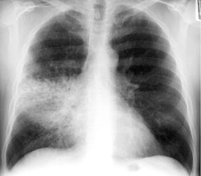
Air Bronchogram A tubular outline of an airway made visible due to the filling of the surrounding alveoli by fluid or inflammatory exudates. Conditions in which air bronchograms are seen: •  Lung consolidation •  Pulmonary oedema •  Non-obstructive pulmonary atelectasis •  Interstitial disease •  Neoplasm •  Normal expiration

Silhouette Sign

Image
Silhouette Sign •  The loss of the lung/soft tissue interface due to the presence of fluid in the normally air-filled lung •  If an intrathoracic opacity is in anatomic contact with a border, then the opacity will obscure that  border •  Commonly seen with the borders of the heart, aorta, chest wall, and diaphragm

Venous eczema

Image
Venous eczema  occurs as a result of venous hypertension causing fluid to collect in the tissues. The stasis of this fluid in the soft tissues results in activation of the innate immune response and subsequent inflammation. Venous eczema has the following clinical characteristics: ·          Itchy red, blistered and crusted  plaques ; or dry fissured and  scaly   plaques  on one or both lower legs (commonly mistaken for cellulitis) ·          Atrophie Blanche (white irregular scars surrounded by red spots) ·          Orange-brown patches pigmentation due to haemosiderin deposition ·          Lipodermatosclerosis If you’re not sure if the patient has venous eczema, ask “Is it itchy?” Varicose eczema is often intensely pruritic and this is a common indication for intervention.

Grading volume of a murmur

Image
Grading Volume of a Heart Murmurs: The experienced examiner should be able to give the murmur a grade according to its loudness:    Grade 1 – very faint murmur which is only audible during prolonged auscultation Grade 2 – a faint murmur, immediately audible with a stethoscope Grade 3 – loud murmur with NO thrill Grade 4 – loud murmur WITH a thrill Grade 5 – loud murmur WITH a thrill, heard with only half of the stethoscope touching the chest Grade 6 – loud murmur WITH a thrill, heard WITHOUT the stethoscope touching the chest Top tip: Never describe a murmur as grade 1 or 2 in OSCE examinations as it is commonly accepted that only expert cardiologists can detect these murmurs! The grading system can be made easier by remembering that grade 4 and above requires the presence of a thrill.

Easy ACLS

Image
Hi again! This is my another android application which is designed for android mobiles only. This app is designed for doctors and medical students who work in ERs and Face heart emergency situations or for the doctors who take ACLS course and prepared themselves for the exam. American Heart Association, as one of the most reputable international bodies, encourages all doctors to employ algorithms that could help millions of people world over. In this app, we collect those algorithms in an application that every single doctor should have them in their pocket to use in the time of need because life is why. We cover some topic like “Acute Coronary Syndrome”, Bradycardia and Tachycardia with pulse and etc. You can download it HERE from Google Play

Easy Arrhythmias

Image
Hi, I developed another 5 stars app for android mobile regarding the cardiac arrhythmias.  This app is designed to give you a fast and reliable review of Cardiac Arrhythmias, especially when you are in your clinic or Emergency Room and face a patient with arrhythmia to diagnose and manage it properly. This app is recommended for Emergency doctors, Medical officers, medical students, and nurses. You can download it HERE from Google Play Enjoy Max

Ankle-Brachial Pressure Index (ABPI)

The ankle brachial pressure index (ABPI) is an easily measured clinical parameter that is often used to assess the adequacy of blood flow to the lower limbs. The patient should lie flat on a bed when having an ABPI measured. The systolic blood pressure in the brachial artery is measured using a stethoscope and sphygmomanometer. The systolic blood pressure at the ankle is then measured using a sphygmomanometer wrapped around the calf and a Doppler probe positioned over the posterior tibial or dorsalis pedis artery. Cuff pressure is inflated until blood flow to the foot is cut off. Flow is slowly re-established by deflating the cuff, and the pressure at which the first Doppler signal is obtained is noted. After performing these tests, the observer will have two figures – the systolic blood pressures in the brachial artery and at the ankle. The ABPI is calculated simply by finding their ratio. In health, the ratio should be greater than or equal to 1. As the value decreases, symptom...

Non Shock-able Rhythms

Image
Defibrillation will not be helpful to patients with one of these rhythms. Cardiopulmonary resuscitation should be administered and attempts made to reverse the cause of cardiac arrest. Pulseless electrical activity (PEA) (also known as electromechanical dissociation or EMD) The heart rhythm is indistinguishable from a heart rhythm normally compatible with life. Asystole There is no identifiable cardiac electrical activity. It is important to adjust the gain on the ECG monitor to ensure that ‘fine’ ventricular fibrillation is not missed.

Shock-able Rhythms

Image
If one of these rhythms is identified, the priority in treatment is to deliver electricity to the heart using a defibrillator. Ventricular fibrillation (VF) This is identified by the erratic nature of the electrical activity. It is random and unpredictable. It is sometimes classified as being fine or coarse depending on whether the electrical activity is of small (fine) or large (coarse) amplitude. Ventricular tachycardia (VT) This is a broad QRS complex tachycardia that has a very distinctive appearance on an ECG monitor. It is not always associated with cardiac arrest but is always a significant arrhythmia.

Cardiac arrest rhythms

When a patient has a cardiac arrest, the heart stops pumping blood around the body. The electrical activity in the heart does not always stop immediately, however, and the immediate advanced management of patients with cardiac arrest is largely determined by exactly what is happening to the heart rhythm. It is imperative that you are able to identify each of the following rhythms.

Reading an ECG

Image
When interpreting an ECG, always use the following headings: Heart rate Heart rhythm Cardiac axis P waves QRS complexes ST segment Q–T interval T waves

Easy Emergencies

Image
Hi, I am back again. Today I finished my third app after a hard work. I call it "Easy Emergencies".  In this app, I covered the most common topics that a practitioner face in his clinic or in an ER. Subjects like Anaphylaxis, Animal bites, MI, Coma, Diabetic Ketoacidosis and etc. I designed it in a way that you can jump to a topic that you want. For example, if you need to know to manage full partial burnings, just click on the button of management and ignore the rest of the data to save your patient time. Here are some screenshots: You can Download it HERE or scan the barcode below: Cheers Max

Easy ECG

Image
   Hey everyone,    This is another application that I developed the other day. with this app, you can easily read and interpret an ECG in the clinic or at a party 😅(You know people, when they know that you are a doctor, they remind their pains and problems!!). Also with this app, you can review the most important aspect of ECG in a fast way. All doctors, medical students and nurses can download and use it.    Here are some screenshots:    You can download it from Google Play or click Here .      Or you can use your Barcode Scanner: Cheers